Glaucoma
What is Glaucoma?
Glaucoma is a leading cause of blindness in the United States, especially for older people. But loss of sight from glaucoma is often preventable if you begin treatment early.
Glaucoma is a disease of the optic nerve, which carries the images we see to the brain. Glaucoma is caused by the pressure inside the eye. The higher the pressure inside the eye, the greater the chance of damage to the optic nerve.
The optic nerve is made up of a huge number of nerve fibers, like an electric cable containing a huge number of wires. Glaucoma can damage nerve fibers, causing blind spots to develop. Usually people don’t notice these blind areas until much optic nerve damage has already occurred. If the entire nerve is destroyed, blindness results.
Early detection and treatment by your ophthalmologist are the keys to preventing optic nerve damage and blindness from glaucoma.
What Causes Glaucoma?
Clear liquid, called the aqueous humor, circulates inside the eye. A small amount of this fluid is produced constantly, and an equal amount flows out of the eye through a microscopic drainage system. (This liquid is not part of the tears on the outer surface of the eye.) You can think of the flow of aqueous fluid as a sink with the faucet turned on all the time.
If the “drainpipe” gets clogged, water collects in the sink and the sink may overflow. Because the eye is a closed structure, the excess fluid cannot overflow if the drain is clogged. If the drainage area of the eye-called the drainage angle-is blocked, the fluid pressure within the inner eye may increase, which can damage the optic nerve.
What are the different types of glaucoma?
Chronic open-angle glaucoma: This is the most common form of glaucoma in the United States. It occurs as a result of aging. The drainage angle of the eye becomes less efficient with time, and pressure within the eye gradually increases.
If this increased pressure results in optic nerve damage, it is known as chronic open-angle glaucoma. Over 90% of adult glaucoma patients have this type of glaucoma.
Chronic open-angle glaucoma damages vision so gradually and painlessly that you are not aware of trouble until the optic nerve is already badly damaged.
Angle-closure glaucoma: Sometimes the drainage angle of the eye may become completely blocked. It is as though a sheet of paper floating near a drain suddenly drops over the opening and blocks the flow out of the sink. In the eye, the iris (the part that makes eyes blue or brown) may act like the sheet of paper closing off the drainage angle.
When eye pressure builds up suddenly, it is called acute angle-closure glaucoma.
Symptoms may include:
- Blurred vision;
- Severe eye pain;
- Headache;
- Rainbow haloes around lights;
- Nausea and vomiting.
If you have any of these symptoms, call your ophthalmologist immediately. Unless an ophthalmologist treats acute angle-closure glaucoma quickly, blindness can result. Acute angle closure glaucoma is more common in Asian people than in people of European descent; it is rare in people of African descent.
In some patients, glaucoma has features of both the chronic open angle type and the acute angle closure type. This may be called chronic angle closure glaucoma or mixed mechanism glaucoma. It occurs more frequently in people of African and Asian descent.
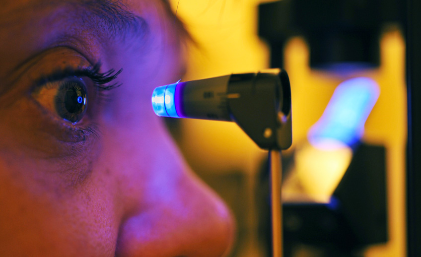
How is glaucoma detected?
Regular eye examinations by our ophthalmologists are the best way to detect glaucoma. During a complete and painless examination, our ophthalmologists will:
- Measure your intraocular pressure (tonometry);
- Inspect the drainage angle of your eye (gonioscopy);
- Evaluate any optic nerve damage (ophthalmoscopy);
- Test the visual field of each eye (perimetry).
Some of these tests may not be necessary for every person. You may need to repeat these tests on a regular basis, to determine if glaucoma damage is increasing over time.
Who is at risk for glaucoma?
High pressure alone does not mean that you have glaucoma. Your ophthalmologist puts together many kinds of information to determine your risk for developing the disease. The most important risk factors include:
- Age;
- Ancestry;
- A family history of glaucoma;
- Past injuries to the eyes.
Our ophthalmologists weigh all of these factors before deciding whether you need treatment for glaucoma, or whether you should be monitored closely as a glaucoma suspect. Being a “glaucoma suspect” means your risk of developing glaucoma is higher than normal, and you need to have regular examinations to detect the early signs of damage to the optic nerve.
How is glaucoma treated?
As a rule, damage caused by glaucoma cannot be reversed. Eye drops, pills, and laser and surgical operations are used to prevent or slow further damage from occurring. With any type of glaucoma, periodic examinations are very important to prevent vision loss. Because glaucoma can worsen without you being aware of it, your treatment may need to be changed over time.
Medicines
Glaucoma is frequently controlled with eye drops taken several times a day, sometimes in combination with pills. These medications decrease eye pressure, either by slowing the production of aqueous fluids within the eye or by improving the flow through the drainage angle.
For these medications to work, you must take them regularly and continuously. It is also important to tell all of your doctors about the eye medications you are using. Glaucoma medications can have side effects. You should notify your ophthalmologist immediately if you think you may be experiencing any side effects.
The side effects from eye drops may include:
- A stinging sensation;
- Red eyes or darkening of the color of the pupil;
- Changes in pulse and heartbeat;
- Changes in energy level;
- Changes in breathing (especially with asthma or emphysema);
- Headaches;
- Blurred vision.
The side effects from pills may include:
- Tingling of fingers and toes;
- Drowsiness;
- Loss of appetite;
- Bowel irregularities;
- Kidney stones;
- Anemia or easy bleeding.
Lasers
You and your doctor may determine that a laser treatment may be a better option for controlling your eye pressure. Lasers such as Selective Laser Trabeculoplasty can do this by clearing debris from the blocked drain. This procedure may be done instead of eyedrops or in addition to the drops you are already taking. SLT treatments are relatively safe and effective for many types of glaucoma and in some cases may be more effective in preventing damage to your vision than drops.
A second type of laser treatment called a Peripheral Iridotomy is used as a “just in case valve” in patients at risk for angle closure glaucoma. Like the hole at the top of the sink in your bathroom, if the water level (pressure) rises due to a blocked drain, a hole can be made in the iris by a laser and can help keep the pressure from shooting up very high and causing permanent damage to the vision.
Minimally Invasive Glaucoma Surgery (MIGS)
This is a group of treatments that are surgical in nature but are generally safer with a quicker recovery than standard glaucoma surgery. If you have mild to moderate glaucoma and a visually significant cataract, a small stent inserted at the time of cataract surgery such as the iStent trabecular microbypass stent may help to allow fluid to drain out of the eye better, partially bypassing the blockage that may exist in the trabecular meshwork.[HYPERLINK TO ISTENT ARTICLE]
Trabeculectomy and Tube Shunts
Designed for more significant glaucoma, these surgeries can more dramatically lower the pressure than any of the above treatments.
Though serious complications of modern glaucoma surgery are rare, they can occur, as with any surgery. Surgery is recommended if our ophthalmologists feel that it is safer to operate than to allow optic nerve damage to continue.
What is your part in treatment?
Treatment for glaucoma requires teamwork between you and your doctor. Our ophthalmologists can prescribe treatment for glaucoma, but only you can make sure you take your eye drops or pills. Never stop taking or change your medications without first consulting your ophthalmologist. Frequent eye examinations and tests are critical to monitor your eyes for any changes. Remember, it is your vision, and you must do your part to maintain it.
Loss of vision can be prevented
Regular medical eye exams may help prevent unnecessary vision loss. Recommended intervals for eye exams are:
- Age 18-39: every 2-3 years;
- Age 40 and over: every 1 to 2 years.