Retinal Detachment and Tears
What is the retina
The retina is the layer of nerve tissue along the back surface of the eyeball that receives light focused by the front of the eye. The retina then sends nerve impulses back to the brain through the optic nerve, where it is interpreted by the visual cortex as an image. Because the retina is nerve tissue, like the brain, any damage to it can result in permanent vision loss.
Treatments
Posterior Vitreous Detachments (PVD)
In this situation, there is no actual tear in the retina itself. Rather, due to age-related liquefaction of the vitreous, the vitreous can physically pull on the outer surface of the retina. When the retina is mechanically-stimulated in this way, it sends impulses to the brain, which interprets the impulses as “flashes” of light. These flashes may be minimal (ie. split-second, peripheral lightning streak) or be extensive (multiple flashes around the eye over hours). “Floaters” are also associated with PVDs, and can be in many different forms, from a large cloudy veil, to multiple dots that zip across one’s vision. Because the acute onset of flashes and floaters can also represent an actual retinal tear or detachment, expedient examination of the retina is required to accurately diagnose the situation. If a PVD is diagnosed, there is no need for treatment, but followup visits are scheduled to ensure that a retinal tear does not develop subsequently.
Retinal Tears
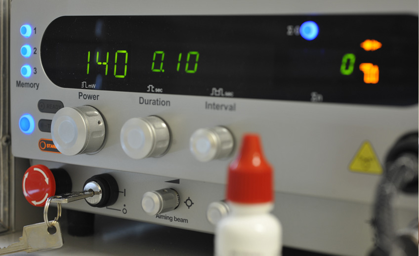
Tears of the retina can occur in any part of the retina, and involve any quadrant of vision. An acute onset of flashes and floaters can represent a retinal tear. In contrast, retinal tears are also sometimes diagnosed on routine examinations of the eye, without any symptoms experienced by the patient! One of the more concerning symptoms of a possible retinal tear is the presence of a shadow, or a piece of the vision that appears to be “missing”. Any such symptoms, particularly in conjunction with flashes and/ or floaters, requires an expedient call to an ophthalmologist or retinal specialist for an evaluation. Depending on the size of the tear, treatment options include laser treatment, cryotherapy, pneumatic retinopexy, or scleral buckle surgery.
Retinal Detachments
Detachments occur when the retina suffers tear, and fluid infiltrates sub-retinally to detach the retina from the back of the eye. Because detachments can result permanent loss of vision, especially when the center of the retina, the macula, is involved. When the macula is threatened by a detachment, surgery is usually indicated to prevent blindness. Unfortunately, even with successful surgery, there is no guarantee that vision can be retained or recovered.